Prompt
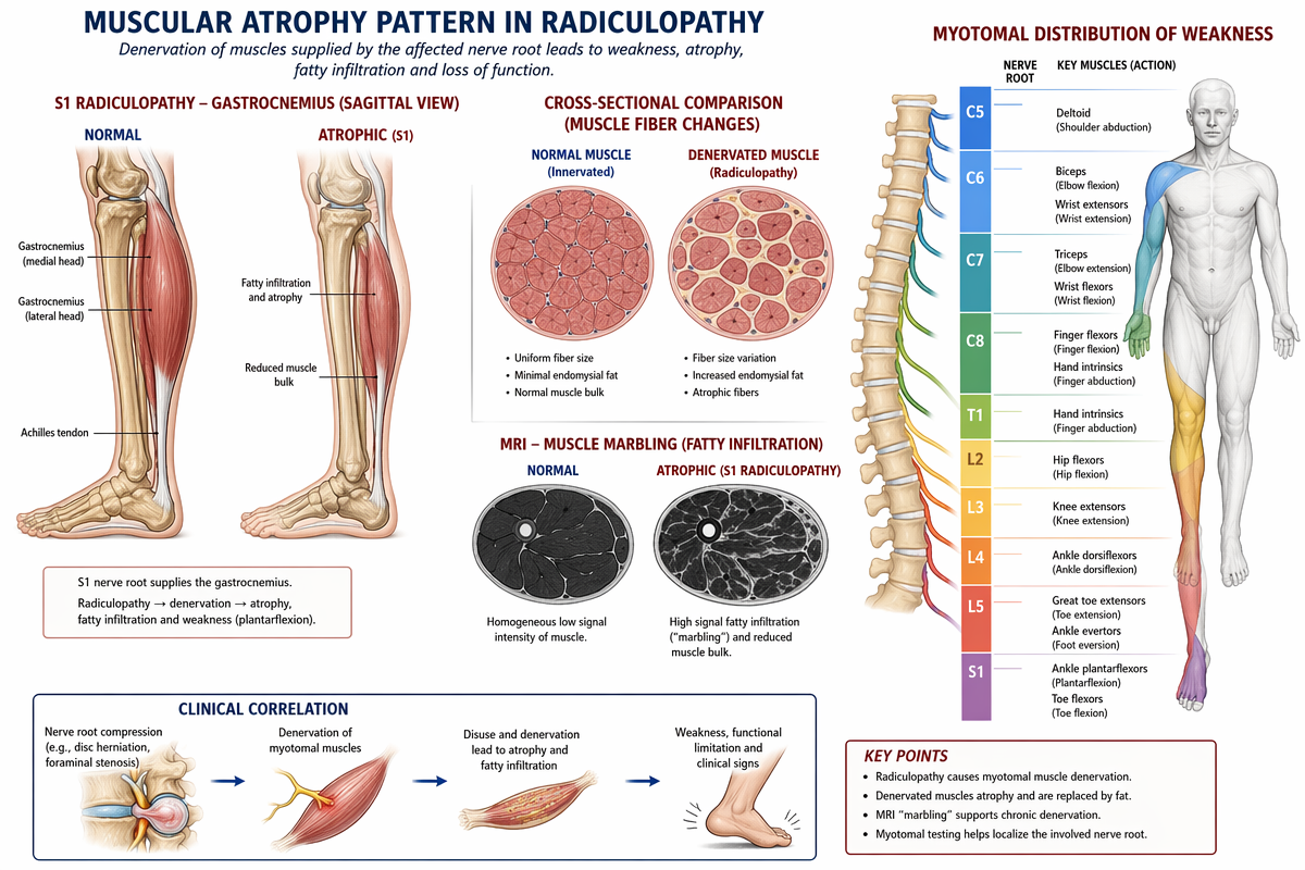
Medical anatomy illustration showing muscular atrophy patterns in radiculopathy. The layout includes a sagittal view comparison of normal vs atrophic gastrocnemius muscle (S1 level), a cross-sectional comparison of muscle fiber denervation, and a marbling pattern on MRI. Includes a myotomal distribution chart of weakness for nerve roots C5-S1 with a human figure diagram. Clinical illustration style, high contrast, white background, educational quality, clean typography.