Prompt
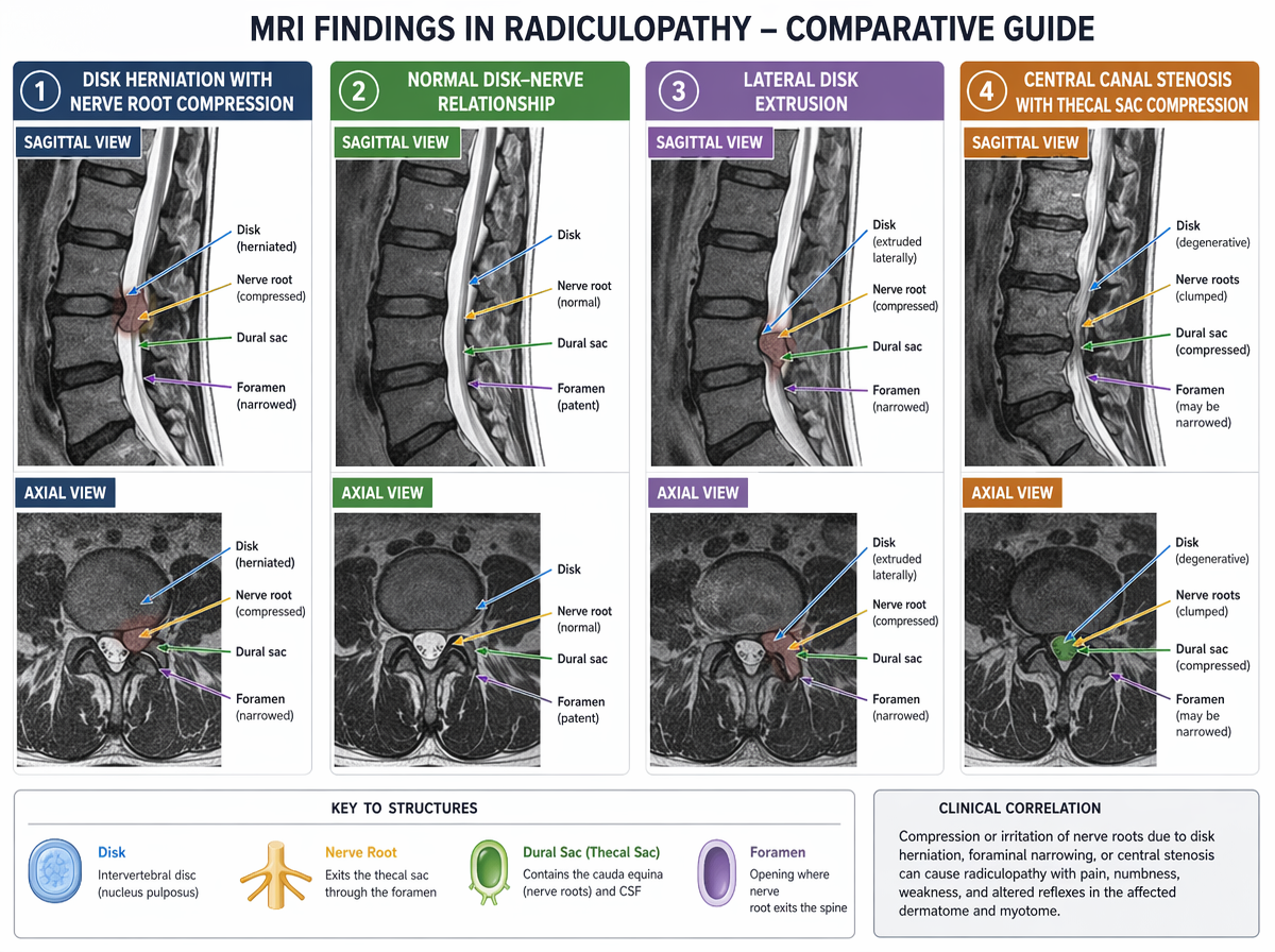
Medical imaging comparison infographic: Side-by-side illustration of MRI findings in radiculopathy. Four columns showing: (1) Disk herniation with nerve root compression, (2) Normal disk-nerve relationship, (3) Lateral disk extrusion, (4) Central canal stenosis. Each column includes sagittal and axial views. Clinical illustration style, high contrast, white background, anatomy-focused. Labels for disk, nerve root, dural sac, and foramen with a color-coded key at the bottom.